If you are wondering how cell imaging in drug discovery works, the short answer is this: automated microscopes photograph cells treated with compounds, and software turns those pictures into measurements that can hint at what a compound does.
This guide walks through the idea in plain language and shows where the figures come from.
| Item | Value |
|---|---|
| Dyes multiplexed in the Cell Painting assay | Six fluorescent dyes |
| Imaging channels used | Five channels |
| Cellular components or organelles revealed | Eight |
| Morphological features measured per cell | About 1,500 |
These figures come from the 2016 Cell Painting protocol in Nature Protocols. Protocols get updated and numbers can change, so check the source before you rely on them.
What cell imaging adds to drug discovery
Drug discovery is a long chain of steps, from finding a biological target to testing candidates in people. If you want the wider map, start with what is drug discovery, then come back to see where microscopy fits.
Imaging is mostly used early on, when teams need to know whether a compound changes living cells in a useful way. A 2007 review in the British Journal of Pharmacology describes how the emergence of automated cellular systems allows rapid visualization of large groups of cells and phenotypic analysis in a quantitative manner.
The key idea is that a picture holds far more information than a single number. A cell can shrink, swell, change its nucleus, rearrange its skeleton or lose organelles, and a microscope can capture all of that at once.
How high-content imaging works step by step
The workflow is similar across laboratories, though details differ by project. In outline:
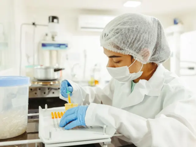
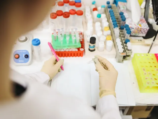
- Plate the cells. Cells are grown in multi-well plates, often with a different compound or genetic change in each well.
- Stain them. Fluorescent dyes or tagged proteins make structures such as nuclei visible.
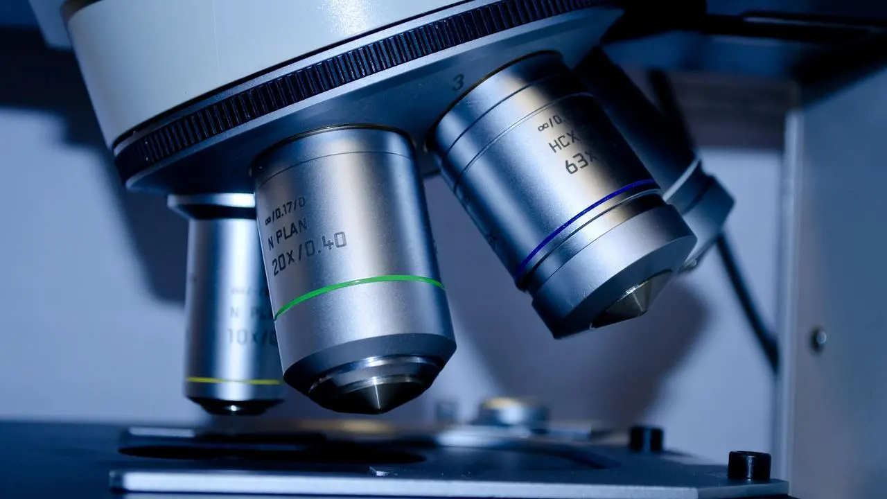
- Image automatically. A robotic microscope photographs each well, usually in several colour channels.
- Segment and measure. Software finds each cell and records size, shape, texture and intensity.
- Compare. Profiles from treated wells are compared with untreated controls and with each other.
Because the plates are processed by machines, this approach pairs naturally with high throughput screening, where very large numbers of compounds are tested in sequence. The difference is that each well yields a rich image rather than a single readout.
Cell Painting: a standard way to profile cells
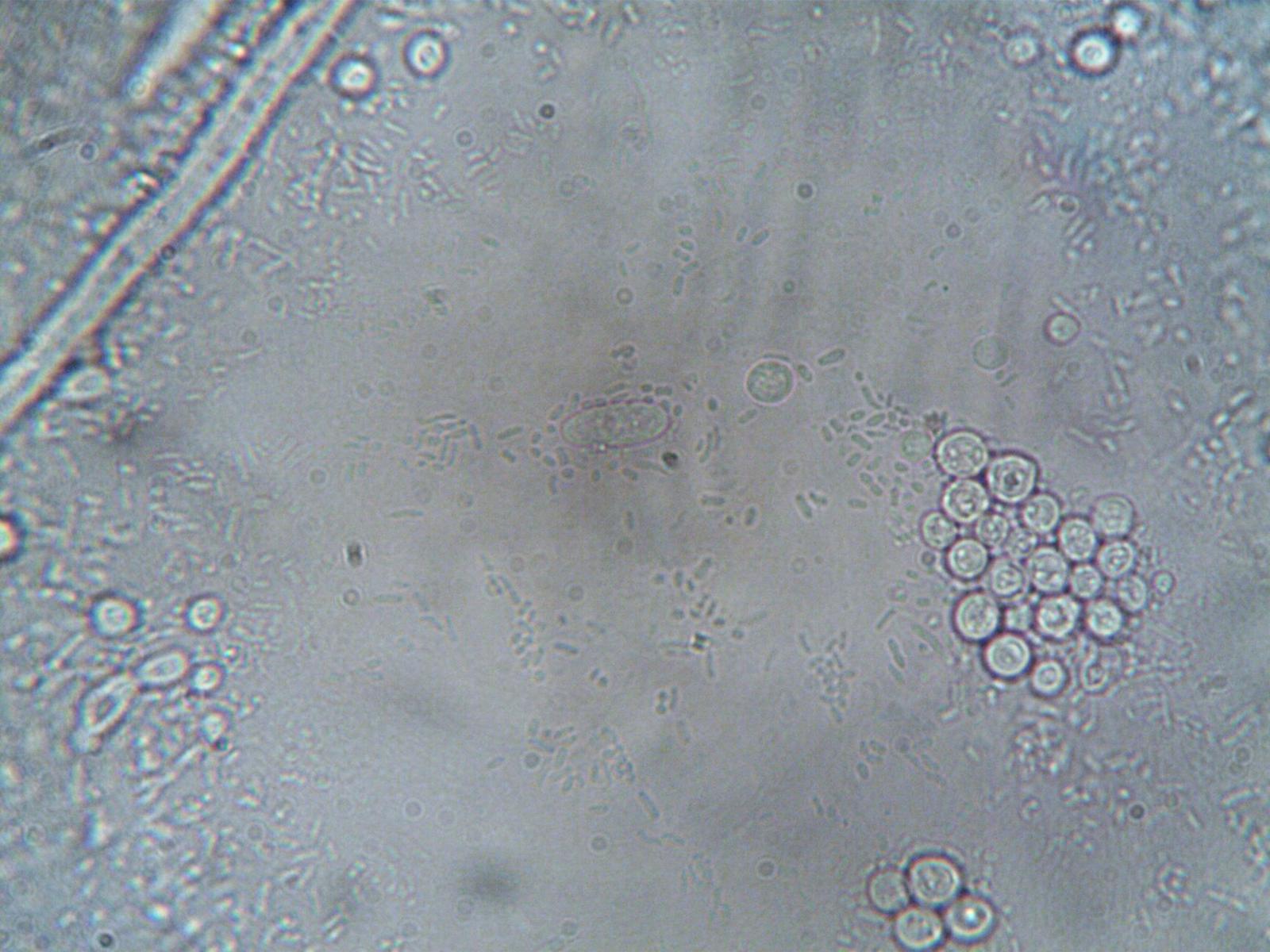
One widely used format is the Cell Painting assay. According to the protocol, it multiplexes six fluorescent dyes, imaged in five channels, to reveal eight broadly relevant cellular components or organelles.
Image analysis software then identifies single cells and, in the protocol’s words, measures ∼1,500 morphological features (various measures of size, shape, texture, intensity, and so on) to produce a rich profile that is suitable for the detection of subtle phenotypes.
Where artificial intelligence enters the picture
Manual inspection cannot keep up with the volume of images these screens produce. A 2024 review in Current Opinion in Structural Biology says that AI’s role in analysis of HCI data from fixed and live-cell imaging is enabling label-free and multi-channel fluorescent screening methods and improving compound profiling.
The same review notes that HCI experiments are rapid and cost-effective, facilitating large data set accumulation for AI model training.
Limits and uncertainty
Imaging does not tell you whether a compound will work in people. Cells in a dish lack the immune system, blood flow and metabolism of a patient, which is one reason many candidates still stumble later; see why do drugs fail in clinical trials for that wider story.
The AI review also cautions that the success of AI in drug discovery also depends on high-quality data, reproducible experiments, and robust validation to ensure model performance. It lists challenges such as the need for annotated compounds and managing vast image data.
Other practical limits are worth knowing:
- Batch effects: plates run on different days or instruments can look different for reasons unrelated to biology.
- Model dependence: results from one cell type may not carry over to another.
- Interpretation: a changed shape shows that something happened, not why.
- Data volume: storing and processing images demands substantial computing resources.
Fixed, live and label-free imaging
Imaging comes in several flavours, and each answers a different question. The AI review above refers to fixed and live-cell imaging, which differ in important ways.
- Fixed-cell imaging freezes cells at one moment and allows many dyes, but it gives only a snapshot.
- Live-cell imaging follows the same cells over time, which helps when a response is gradual or short-lived.
- Label-free imaging avoids dyes altogether and relies on software to pick out structures.
Choosing among them is a trade-off between detail, time resolution, cost and the risk that the dye itself disturbs the cells. No single format is best for every project.
Reading an imaging result sensibly
Teams usually look at whether a profile is reproducible across replicate wells, whether it changes smoothly with dose, and whether it is distinct from general toxicity.
A compound that simply kills cells can look dramatic under the microscope. Careful design therefore includes counting cells and checking that the profile reflects something more specific than damage.
Researchers also compare image-based hits with other kinds of evidence, such as biochemical assays on the suspected target. Agreement between independent methods raises confidence, while disagreement is a reason to look harder rather than to pick the more appealing result.
Who uses these methods and why it matters to you
Academic laboratories, public consortia and pharmaceutical teams all use image-based profiling.
For a general reader, the useful takeaway is that imaging is a tool for narrowing choices, not a verdict on safety or benefit. Nothing here is medical advice, and questions about any treatment belong with a physician or other qualified health professional.
Imaging results can inform which ideas go forward, but they sit many steps before any decision about use in people.
Frequently asked questions
What is high-content imaging?
Show answer
It is automated microscopy combined with quantitative image analysis, used to measure many features of cells after they are exposed to compounds or genetic changes.
Does cell imaging show that a drug works?
Show answer
No. It can show that a compound changes cells in a particular way. Whether that matters in a patient has to be tested in later studies, and any personal health question should go to a physician.
What does Cell Painting measure?
Show answer
Per the 2016 protocol, it uses six dyes in five channels to reveal eight cellular components, and software extracts about 1,500 features per cell.
Why is AI used with microscopy images?
Show answer
Screens generate far more images than people can review. AI can help analyse imaging data and improve compound profiling, though it depends on good data and validation.
This article is for information only. It is general education about science and the drug industry, and it is not medical, treatment or investment advice.